
Improving detection of breast cancer
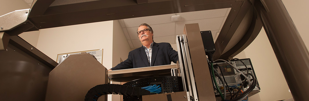
Dr. John Boone is a recognized expert in the field of medical imaging, with a focus on improving breast cancer detection. He and his team have developed a device with the potential to detect tumors in the breast earlier and with less discomfort.
The American Cancer Society reports that breast cancer is the second most common form of cancer among American women, following skin cancers. It estimates that about 1 in 8 women in the U.S. will develop invasive breast cancer during her lifetime.
Traditionally, mammograms have been used to detect breast cancers as part of a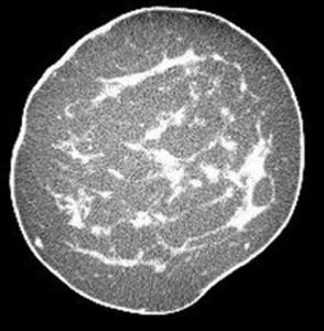 regular screening, but Boone has developed what could be a better approach, hopefully improving both detection and patient outcomes. Boone and his team have designed and developed an innovative computed tomography (CT) scanner designed specifically for imaging the breast (UC Case 2005-543). The intended advantage of this device is that it provides a true three-dimensional, highly-detailed image of the human breast, offering a less obstructed view of potential lesions than the current two-dimensional mammogram.
Unlike mammography, the scanner does not require compression of the breast. Instead, the patient lies face down on a padded table and places the breast in a circular opening. The scanner generates 300 to 500 tomographic image “slices” of the breast, which are then assembled into a three-dimensional digital model. The imaging procedure takes approximately 10 seconds.
Thanks to support from the National Institutes of Health, Boone’s team has assembled four scanners that have been used to image over 600 women at the UC Davis Medical Center.
The technology led to the formation of Isotropic Imaging Corporation, which intends to license the technology developed at UC Davis to commercialize a scanner.